
Estimation of Sex by Pattern of Calcification of First Costal Cartilage in North Indian Population
Article Sidebar
Main Article Content
Abstract
Objective- Estimation of sex by pattern of calcification of first costal cartilage in North Indian Population.
Methods- The present study has been conducted in the “Department of Anatomy in collaboration with the Department of Forensic Medicine and Department of Radiology,PGIMS, Rohtak”.
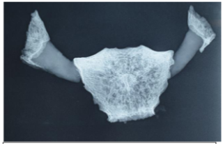
The specimen consisted of 50 pairs of first ribs and manubrium from both sexes, aged between 15 and 30 years, in order to determine the pattern of calcification. Specimens were taken from “cases of medico-legal autopsies” performed in the “Department of Forensic Medicine” with the legal heir of the deceased's consent, and they were radiographed in the “Department of Radiology”.
Results- In the present study, calcification was observed in cartilages of both sexes aged above 16 years. Type A (marginal bracket), type A 1 (marginal linear) and type B (central) pattern were found more frequently in females on both sides with an estimated predictive value of 66.32%, 50.27% and 100% respectively. Type C (mixed) pattern occurred more frequently in males on both sides with estimated predictive value to be 100%.
Conclusion- There was no evidence of bilateral asymmetry between the calcification patterns on the right and left sides. This means that the first costal cartilage's calcification displays a clear pattern in respect to the sex at issue.
Article Details
References
Standring S. Gray’s Anatomy. 41st ed. London: Elsevier Limited; 2016.
Cunningham DJ. Cunningham’s manual of practical Anatomy. 15th ed. New York: Oxford University press; 2008.
Datta AK. Essentials of Human Anatomy. 9th ed. Kolkata: Current Books International; 2014.
King JB. Calcification of costal cartilages. Br J Radiol. 1939; 12(133): 2-12.
Vastine JH. Genetic influence of osseous development with particular reference to the deposition of calcium in the costal cartilages. American J Roentgenol. 1948; 59(2): 213-21.
Ray K, Bardhan J, Sarkar KN. A study of calcification of costal cartilages (1st To 7th) in different age groups and its effect on chest expansion in both male and female. J dental Med Sci. 2017; 16(3): 115-23
Rao NG, Pai LM. Costal cartilage calcification pattern-a clue for establishing sex identity. Forensic Sci Int. 1988; 38(3-4): 193-202.
Sanders CF. Sexing by costal cartilage calcification. Br J Radiolog. 1966; 39(459): 233.
Elkeles A. Sex differences in the calcification of costal cartilages. J Am Geriatr Soc. 1966; 14(5): 456-62.
Navani S, Shah JR, Levy SP. Determination of sex by costal cartilage calcification. Am J Roentogenol Radiother. 1970; 108(4): 771-4.
Zhang S, Zhen J, Li H, Sun S, Wu H, Shen P, Chen Z, Yang C. Characteristics of Chinese Costal Cartilage and Costal Calcification Using Dual-Energy Computed Tomography Imaging. Sci Rep. 2017; 7(1): 1-10.
Inoi T. Estimation of sex and age by calcification pattern of costal cartilage in Japenese. Nihon Hoigaku Zaashi. 1997; 51(2): 89-94.
Vastine JH. Genetic influence of osseous development with particular reference to the deposition of calcium in the costal cartilages. American J Roentgenol. 1948; 59(2): 213-21.
Ray K, Bardhan J, Sarkar KN. A study of calcification of costal cartilages (1st To 7th) in different age groups and its effect on chest expansion in both male and female. J dental Med Sci. 2017; 16(3): 115-23.
Raghvendra DR, Nirmala D. Multiple ossified costal cartilages for 1st rib. Int J Anat Res. 2014; 2(4): 744-47.