
A Cross-Sectional Study Using MR Imaging To Evaluate Cerebral Venous Thrombosis
Article Sidebar
Main Article Content
Abstract
Background:Dural sinus thrombosis, along with the thrombosis of the deep or cortical cerebral venous system and the venous stroke resulting from it, is more common than once thought. Cerebral venous thrombosis (CVT) is a cause of stroke with diverse etiologies and varied clinical presentations. Its manifestations may simulate an acute arterial stroke or a mass lesion. The pathophysiology of CVT with associated venous strokes appears to differ from arterial strokes. Acute arterial strokes show cytotoxic edema, whereas venous strokes are thought to contain vasogenic and interstitial edoema due to venous congestion. MR imaging has been deemed to play a crucial role in diagnosing and evaluating this complex disease and, hence, has been progressively used to guide and direct its management.
Purpose:To study the various aspects of cerebral venous thrombosis in conventional and advanced MR imaging sequences
Methodology:This is a prospective observational study of 50 patients with cerebral venous thrombosis (CVT). Patients had undergone conventional MRI, diffusion-weighted imaging, and MR venogram. The diagnosis of CVT was confirmed with an MR venogram and other conventional MR sequences in 48 patients. MR contrast venography was done in 2 patients.
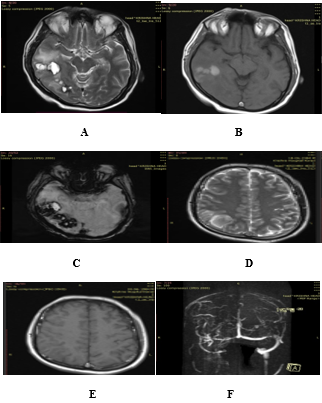
Result:In our study, headache was the most common symptom in 38/50 patients (76%), followed by a focal neurological deficit in 19/50 (38%), seizures, and vomiting in 13/50 patients (26% of each). In our study, the most common site of sinus thrombosis was found to be the superior sagittal sinus (70%), followed by the transverse and sigmoid sinuses (42% and 22%, respectively). The involvement of the deep venous sinus was only present in three (6%) patients. The sinus thrombus clot age was found to be in sync with clinical presentation and parenchymal imaging findings in 90% of cases, whereas in 10% of cases the sinus thrombus age was older than the parenchymal findings. Hemorrhagic infarct was identified in 40% of sinus thrombosis patients, intraparenchymal hematoma in 16% of patients, and non-hemorrhagic infarct in 38% of patients. In the acute phase, it was seen that in our study, most patients (7 patients) presented with a non-hemorrhagic infarct, whereas in the subacute phase, 18 cases were of hemorrhagic venous infarct. There was a significant correlation (with p-values less than 0.05) between the presence of hemorrhagic infarction in the subacute phase and intraparenchymal hematoma in the acute phase.
Conclusion:Evaluation of CVT is often a difficult task, but after the introduction of newer MR imaging techniques, it is possible to predict if brain lesions, detected clinically and using conventional MR imaging methods, may lead to full recovery, as expected in arterial infarcts and even a hematoma. Our research concludes that MRI is the best tool to evaluate CVT.
Article Details
References
Lovblad, Karl-Olof a; Bassetti, Claudio b, Schneider, Jacques A,Diffusion-Weighted MR in Cerebral Venous Thrombosis: Cerebrovascular Diseases2001;11(3):169-176,
Bousser MG. Cerebral venous thrombosis: diagnosis and management. J Neurol2000; 247:252–8.
J.C.Corvol,MD,C.Oppenheim,MD,R.Manai,MD;M.Logak,Diffusion-weightedmagneticresonanceimaginginacaseofcerebralvenousthrombosis. Stroke.1998;29:2649-2652.
Bakshi R, Lindsay BD, Bates VE, et al. cerebral venous infarctions presenting as enhancing space-occupying lesions: MR findings. J Neuroimaging 1998; 8:210-215.
Manzione J, Newman GC, Shapiro A, Santo-Ocampo R: Diffusion- and perfusion-weighted MR imaging of dural sinus thrombosis. Am J Neuroradiol 2000; 21:68-73.
María Canedo-Antelo, Sandra Baleato-González, Antonio J. Mosqueira, Jéssica Casas-Martínez et al. Radiologic Clues to Cerebral Venous Thrombosis RadioGraphics 2019; 39:1611–1628
Gates PC. Cerebralvenous thrombosis. A retrospective review. Aust N Z J Med 1986;16:766-70.
Vandenbroucke JP, Rosing J, Bloemenkamp KW, Middeldorp S, Helmerhorst FM, Bouma BN, et al. Oral contraceptives and the risk of venous thrombosis. N Engl J Med 2001;344:1527-35.
Martinelli I, Sacchi E, Landi G, Taioli E, Duca F, Mannucci PM. High risk of cerebral-vein thrombosis in carriers of a prothrombingene mutation and in users of oral contraceptives. N Engl J Med1998;338:1793-7.
Uzan M, Ciplak N, Dashti SG, Bozkus H, Erdinçler P, Akman C. Depressed skull fracture overlying the superior sagittal sinus as a cause of benign intracranial hypertension. Case report. J Neurosurg1998;88:598-600.
Allroggen H, Abbott RJ. Cerebral venous sinus thrombosis. Postgrad Med J 2000;76:12-5.
Karthikeyan D, Vijay S, Kumar T, Kanth L. Cerebral venous thrombosis-spectrum of CT findings. Ind J Radiol2004;14:129-37.
Greiner FG, Takhtani D. Neuroradiologycase of the day. Superior sagittal sinus thrombosis and infarcts. Radiographics1999;19:1098-101.
Bousser MG, Russell RR. Cerebral venous thrombosis. Philadelphia. WB Saunders;1997. p. 385-9.
Leach JL, Bulas RV, Ernst RJ, et al. MR imaging of isolated cortical vein thrombosis: the hyperintense vein sign. J NeuroVasc Dis. 1996;1(3):1-7.
Kumral E, Polat F, Uzunköprü C, Callı C, Kitiş Ö. The clinical spectrum of intracerebral hematoma, hemorrhagic infarct, non-hemorrhagic infarct, and non-lesional venous stroke in patients with cerebral sinus-venous thrombosis. Eur J Neurol. 2012;19:537–543. doi: 10.1111/j.1468-1331.2011.03562.x.
Tsai FY, Wang AM, Matovich VB, Lavin M, Berberian B, Simonson TM, et al. MR staging of acute dural sinus thrombosis: Correlation with venous pressure measurements and implications for treatment and prognosis. AJNR Am J Neuroradiol1995;16:1021-9.
James L, Leach MD, et al. Imaging of cerebral venous thrombosis: current techniques, a spectrum of findings, and diagnostic pitfalls. Radiographics 2006;26:S19- S43
Kon Chu, MD; Dong-Wha Kang, MD, Ph.D.; Byung-Woo Yoon, MD, Ph.D.; Jae- KyuRoh, MD, Ph.D.: Diffusion-weighted magnetic resonance in cerebral venousthrombosis: Arch Neurol. 2001; 58:1569-1576.
James L, Leach MD, et al. Imaging of cerebral venous thrombosis: current techniques, the spectrum of findings, and diagnostic pitfalls. Radiographics 2006;26:S19- S43
Atlas SW, Dubois P, Singer MB, Lu D. Diffusion measurements in intracranial hematomas: implications for MR imaging of acute stroke. AJNR Am J Neuroradiol. 2000;21:1190-1194.
Pascal Favrole MD, Jean-Pierre Guichar MD, et al. Diffusion-Weighted Imaging of Intravascular Clots in Cerebral Venous Thrombosis. Stroke 2004;35:99-103.