
Assessment of Correlation of Meiboscale with Symptom Score Index and Meibomian Gland Dysfunction Sign Score
Article Sidebar
Main Article Content
Abstract
Background:The most frequent mechanism for MGD is a low delivery state marked by gland obstruction. The present study was conducted to assess association of Meiboscale with symptom score and meibomian gland dysfunction sign score.
Materials & Methods: sixty subjects having meibomian gland malfunction of both sexes underwent a complete visual assessment. The MGD sign value was estimated in both eyes through the sum of 6 grading systems. Association among OSDI score, sign scoreand MGL score based on Meiboscale was evaluated.
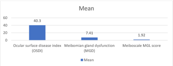
Results: Out of sixty subjects, men were 38 whereas women were 22. The average ocular surface disease index (OSDI) was 40.3, Meibomian gland dysfunction (MGD) was 7.41 and Meiboscale MGL score was 1.92. There was strong correlation of MGL score with MGD sign score (P< 0.05). There was no correlation between MGL and OSDI and OSDI with MGD sign score (P> 0.05).
Conclusion: Meiboscale can be used for reliable assessment and grading ofMGD, and has clinical utility similar to the sum of six MGD sign scores.
Article Details
References
Pult H, Riede Pult BH, Nichols JJ. Relation between upper and lower lids’ meibomian gland morphology, tear film, and dry eye. Optom Vis Sci 2012;89:E310 5.
Weng H Y, Ho W T, Chiu C Y, Tsai T Y, Chang S W. Characteristics of tear film lipid layer inyoung dry eye patients. J Formosan Med Assoc 2021;120:1478 84.
Arita R, Itoh K, Inoue K, Amano S. Noncontact infrared meibography to document age related changes of the meibomian glands in a normal population. Ophthalmology 2008;115:911 5.
Yokoi N, KomuroA, Yamada H, Maruyama K, Kinoshita S. A newly developed video meibography system featuring a newly designed probe. Jpn J Ophthalmol2007;51:53 6.
Pult H, Nichols JJ. A review of meibography. Optom Vis Sci 2012;89:760 9.
Matsuoka T, Tsumura T, Ueda H, Hasegawa E . Video meibographic observations of the meibomian gland. RinshoGanka1996;50:351 4.
Pult H. Relationships between meibomian gland loss and age, sex, and dry eye. Eye Contact Lens 2018;44(Suppl 2):S318 24.
Robin M, Liang H, Rabut G, Augstburger E, Baudouin C, Labbé A. The role of meibography in the diagnosis of meibomian gland dysfunction in ocular surface diseases. Transl Vis Sci Technol2019;8:6.
Brooks CC, Gupta PK. Meibomian gland morphology among patients presenting for refractive surgery evaluation. Clin Ophthalmol2021;15:315 21.
Nichols KK, Foulks GN, Bron AJ, et al. The international workshop on meibomian gland dysfunction: executive summary. Investigative Ophthalmology & Visual Science. 2011; 52(4):1922– 1929.
Guarnieri A, Carnero E, Bleau AM, Alfonso Bartolozzi B, Moreno Montañés J. Relationship between OSDI questionnaire and ocular surface changes in glaucomatous patients. Int Ophthalmol2020;40:741 51.
Foulks GN, Bron AJ. Meibomian gland dysfunction: a clinical scheme for description, diagnosis, classification, and grading. The Ocular Surface. 2003; 1(3):107–126.
Korb DR, Henriquez AS. Meibomian gland dysfunction and contact lens intolerance. Journal of the American Optometric Association. 1980; 51(3):243–251.
Lemp MA, Crews LA, Bron AJ, et al. Distribution of aqueous-deficient and evaporative dry eye in a clinic-based patient cohort: a retrospective study. Cornea. 2012; 31(5):472–478.
Nishant P, Ramawat A, Shrinkhal N, Gupta N, Mittal SK. Correlation of meiboscale, symptom score and sign score for primary meibomian gland dysfunction in Indian eyes – A cross sectional study. Indian J Ophthalmol2022;70:1958-62