
Histo Morphological Evaluation of Esophageal Lesions
Article Sidebar
Main Article Content
Abstract
Background: Esophagus is a tubular structure of the gastrointestinal tract with primary pathological lesions including neoplastic and non-neoplastic conditions. It can secondarily be involved by mediastinal lesions, hence the need to detect and differentiate lesions at an early stage.
This study has been incorporated to emphasize the prevalence of different esophageal lesions with the site-wise distribution along with the comparison of their incidences to age and sex.
Methodology: Our study was done at the department of Pathology, father muller medical college, in Mangalore from January 2020 to January 2021. All the cases were assessed concerning age, chief complaints, site of lesion, and histomorphology. Hematoxylin and eosin staining was performed on paraffin-embedded sections which were microscopically analyzed and categorized into various categories.
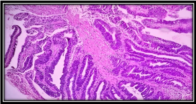
Results: The study included a total of 61 cases with various ages ranging from 31 to 80 years and a mean age presentation of 52.5 years. The most common primary chief complaint was dysphagia 70%. Neoplastic conditions constituted 78.7 % with the most common neoplastic condition being Squamous cell carcinoma at 67.7%
Conclusions: In the present study, we demonstrate that there can be different kinds of lesions in the esophagus all presenting with a similar complaint of dysphagia. Hence it is necessary to evaluate the microscopic findings of each case with due diligence. A spectrum of various Inflammatory and neoplastic conditions can be seen to occur in the esophagus.
Article Details
References
Sung, H, Ferlay, J, Siegel, RL, Laversanne, M, Soerjomataram, I, Jemal, A, Bray F. Global cancer statistics 2020: GLOBOCAN estimates of incidence and mortality worldwide for 36 cancers in 185 countries. CA Cancer J Clin. 2021: 71: 209- 249. https://doi.org/10.3322/caac.21660 .
Choong C, Meyers B. Benign esophageal tumors: introduction, incidence, classification, and clinical features. Seminars Thorac Cardiovasc Surg. 2003; 15:3–8.
Lewis RB, Mehrotra AK, Rodriguez P, Levine MS. From the radiologic pathology archives: esophageal neoplasms: radiologic-pathologic correlation. Radiographics. 2013;33:1083–1108.
Napier KJ, Scheerer M, Misra S. Esophageal cancer: A Review of epidemiology, pathogenesis, staging workup and treatment modalities. World J Gastrointest Oncol. 2014;6(5):112-120. doi:10.4251/wjgo.v6.i5.112
Postlethwait RW. Carcinoma of the thoracic esophagus. Surg Clin North Am. 1983;63:933–940.
Orringer, M. B., Marshall, B., & Iannettoni, M. D. (1999). Transhiatal esophagectomy: clinical experience and refinements. Annals of surgery, 230(3), 392–403.
Choksi D, Kolhe KM, Ingle M, et al. Esophageal carcinoma: An epidemiological analysis and study of the time trends over the last 20 years from a single center in India. J Family Med Prim Care. 2020;9(3):1695-1699. Published 2020 Mar 26. doi:10.4103/jfmpc.jfmpc_1111_19
Bukhari U, Siyal R, Memon F, Memon J. Oesophageal carcinoma: A review of endoscopic biopsies. Pak J Med Sci 2009;25(5):845-848
Kavitha Y, Anjana M.L Histopathological Spectrum of Upper Gastrointestinal Endoscopic Biopsies in a Tertiary care centre Annals of Pathology and Laboratory Medicine, Vol. 8, Issue 6, June 2021
Martin L, Senesse P, Gioulbasanis I, Antoun S, Bozzetti F, Deans C, et al. Diagnostic criteria for the classification of cancer-associated weight loss. VEJ Clin Oncol. 2015 Jan 1; 33(1):90-9
Deans DA, Tan BH, Wigmore SJ, et al. The influence of systemic inflammation, dietary intake and stage of disease on rate of weight loss in patients with gastro-oesophageal cancer. Br J Cancer. 2009;100(1):63-69. doi:10.1038/sj.bjc.6604828
Krishnappa R, Horakerappa MS, Ali K, Gouri M. A study on histopathological spectrum of upper gastrointestinal tract endoscopic biopsies. Int J Med Res Health Sci. 2013;2(3):418-24
Shilpi S, Wani A, Ritika J. Endoscopic biopsies - A boon to diagnose gastrointestinal tract diseases. IAIM, 2019; 6(12): 47-56
Cherian et al. Trends in esophageal carcinoma in Southern India. J Gastrointestin Liver Dis. 2007;16(3):245- 249.
Zhang Y . Epidemiology of esophageal cancer. World J Gastroenterol. 2013 Sep 14; 19(34):5598-606
Fernandes ML, Seow A, Chan YH, Ho KY. Opposing trends in incidence of esophageal squamous cell carcinoma and adenocarcinoma in a multi-ethnic Asian country. Am J Gastroenterol. 2006;101(7):1430-1436. doi:10.1111/j.1572-0241.2006.00570.x
Hansson LE, Sparén P, Nyrén O. Increasing incidence of both major histological types of esophageal carcinomas among men in Sweden. Int J Cancer. 1993;54(3):402-407. doi:10.1002/ijc.2910540309
Das KC, Singh S, Pawar G, Masih R, Raju N. Risk factors analysis of squamous cell carcinoma esophagus in north Indian females in teritiary care hospital: A case-control study. Int J Rcent Sci Res 2015;6:4661- 4
Euchmaur M, Potet F, Goldfain D. Mucin histochemistry of the columnar epithelium of the oesophagus (Barrett’s oesophagus): a prospective biopsy study. J Clin Pathol1984;37:607–10.
Abilash SC, Hasaf K, Gitanjali MM, Shreelaxmidevi S,Balamuruganvelu S. Histopathologic spectrum of upper gastrointestinal tract mucosal biopsies: A retrospective study. Sch. J. App. Med. Sci. 2016;4(5):1807-13.3
Yevoor U, N Ramya, C Swathi. Efficacy of endoscopic biopsies in establishing histologic diagnosis of upper gastrointestinal tract lesions - A one year prospective study. Journal of Diagnostic Pathology and Oncology. 2017 Oct-Dec;2(4):91-94. 24.
Khandelia, R., & Saikia, M. (2017). Histopathologic Spectrum of Upper Gastrointestinal Tract Mucosal Biopsies: A Prospective Study. International Journal of Medical Science and Clinical Invention, 4(11), 3314–3316. https://doi.org/10.18535/ijmsci/v4i11.11
Hirachand, S., Sthapit, R., Gurung, P., Pradhanang, S., Thapa, R., Sedhai, M., & Regmi, S. (2018). Histopathological spectrum of upper gastrointestinal endoscopic biopsies. Journal of BP Koirala Institute of Health Sciences, 1(1), 67–74. https://doi.org/10.3126/jbpkihs.v1i1.19760
Somani N, Patil P. Histopathological Study of The Upper Gastrointestinal Tract Endoscopic Biopsies.Annals of pathology and laboratory medicine august 2018; vol 5 issue 8:683-687